
Smartphone Bedside Fundoscopy
A 3D printed head-mounted smartphone holder to aid physicians in taking portable bedside retinal photographs.
EXPLORE THE PROJECT
Traditional smartphone fundus imaging requires two hands, making it difficult in uncooperative patients.
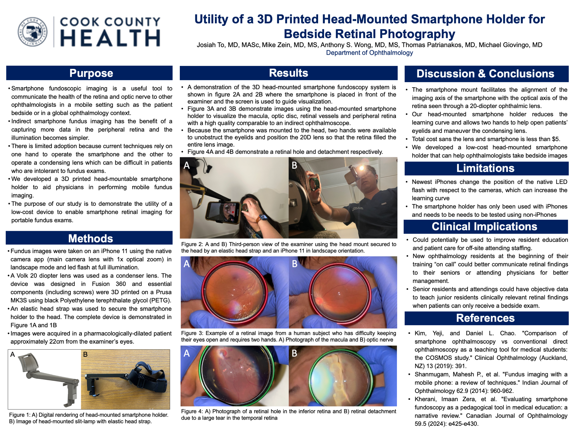
We developed a low-cost, 3D printed head-mounted smartphone holder that enables improved bedside smartphone fundoscopy where the smartphone is suspended in front of the physician’s face so that one hand can be used to open patients’ eyes and the other can hold the 20D condensing lens.
The device allows high-quality fundus photos while freeing both hands to manage eyelids and lens positioning.
Ideal for bedside exams, resident training, and global ophthalmology settings.
PROJECT GOALS
The head-mounted smartphone holder securely positions a smartphone in front of the examiner’s eye, aligning the camera with the optical axis of a standard 20-diopter condensing lens. Once worn, the examiner uses both hands to hold open the patient’s eyelids and adjust the lens position. This setup enables indirect retinal imaging through pharmacologically dilated pupils.
The smartphone’s native camera app (with LED flash at full brightness) is used to capture fundus images in real time. The examiner visualizes and adjusts alignment via the smartphone screen, allowing for quick acquisition of macula, optic nerve, and peripheral retina images.
HOW IT WORKS
The device was designed using Autodesk Fusion 360 and fabricated using a Prusa MK3S 3D printer. All structural components, including adjustable mounting screws, were printed in black PETG plastic for durability and flexibility. A standard elastic head strap was used to secure the device to the examiner’s head.
The imaging system was tested with an iPhone 11 using its primary camera in landscape mode at 1x optical zoom. A Volk 20D lens served as the condensing lens for retinal visualization. The complete unit allows the smartphone to sit approximately 22 cm from the examiner’s eyes—an optimal distance for indirect viewing.
HOW ITS BUILT
Using the head-mounted setup, we captured clear, high-resolution images of key retinal structures—including the optic disc, macula, blood vessels, and peripheral retina. Image quality was comparable to traditional indirect ophthalmoscopy.
Importantly, the head-mounted design enabled bimanual control of the patient’s eyelids and lens positioning, improving image capture efficiency and comfort. We successfully documented retinal pathologies such as retinal holes and detachments using this low-cost system.
RESULTS
Non-diseased Human Retina
Example of a retinal image from a human subject who has difficulty keeping their eyes open and requires two hands. A) Photograph of the macula and B) optic nerve
Pathologic Retina
A) Photograph of a retinal hole in the inferior retina and B) retinal detachment due to a large tear in the temporal retina
FUTURE GOALS
1. Device Development
Testing compatibility with non-iPhone smartphones and additional camera models.
Optimizing the holder design to account for newer iPhone models with different flash-camera configurations.
Evaluating the device in low-resource clinical environments and during on-call ophthalmology training.
Exploring open-source distribution or manufacturing kits to promote global accessibility.
2. Clinical Next Steps
Recruit normal patients to evaluate resident efficacy in using the system.
Teach incoming residents how to use the photography system so they can use it while on-call to gather data and fill out end-user surveys.
PRESENTING THE WORK
We presented present this work at the 2025 Vit-Buckle society meeting in Austin Texas as well as the 2025 internal Cook County Research Day where we won the “Outstanding Moderate Poster Award”.